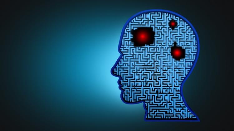
Oxygen maps will help in the treatment of tumours

The amount of oxygen in the tumour affects the success of radiation therapy. The less oxygen, the more difficult the treatment and the higher dose of radiation it requires. Polish researcher is working on oxygen maps that will show which tissues are oxygen-deficient and which have it in abundance.
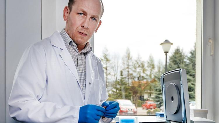
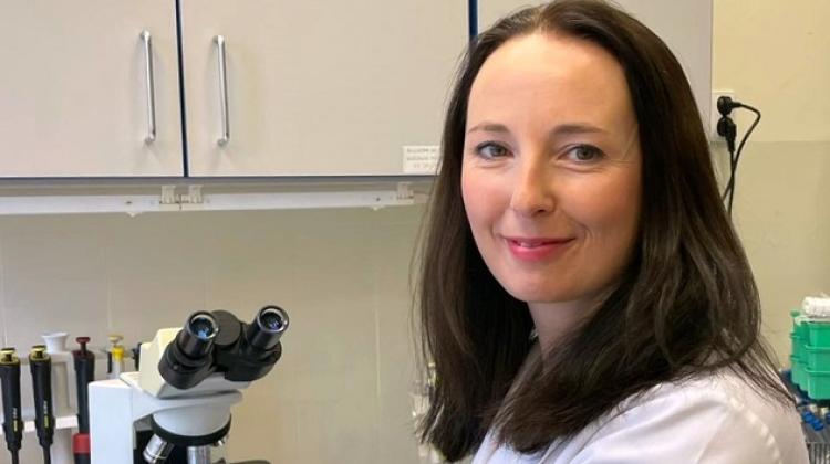
Tumours that form in the human or animal body, have significantly less oxygen compared to healthy tissue. In addition, some points of a tumour can be much better oxygenated than others. "What\'s more important, very large differences in the degree of oxygenation of tumour cells occur between individual patients" - told PAP Dr. Martyna Elas of the Faculty of Biochemistry, Biophysics and Biotechnology, Jagiellonian University.
Her research group has demonstrated that the degree of oxygenation of tumour cells is important in the effectiveness of radiotherapy. "If there is a lot of oxygen in a given cell or tissue, the radiation therapy will be very effective. If the tumour does not have enough oxygen, the cells become resistant to radiotherapy and we must increase the dose of radiation for this patient" - explained the researcher.
But - as the researcher explained - radiotherapeutic treatment today is based on the assumption that the oxygenation of all cells is the same. The reason is the lack of a good method that would allow to determine the oxygenation of tissues and cells in a patient. Oxygen maps developed by Dr. Martyna Elas can help with this situation.
"We want to oxygen maps to be used in clinics and allow for precise and quantitative determination of the concentration of oxygen in the tissues. At the moment it is quite difficult. Oxygen electrodes or fluorescent sensors are currently used for this purpose. Both are needles, which are introduced into a tumour and oxygen concentration is checked at one or more points on the path of the needle. This method is very selective. Therefore, non-invasive preparation of oxygen maps can bring a lot more information" - the researcher described.
Preparation of such a map, however, is quite a difficult, because the animal - and ultimately the patient - must be given a compound, an oxygen probe, which is sensitive to oxygen concentration. It sends a signal which can be observed by means of CT or the spectrometer. By using electron paramagnetic resonance spectroscopy (EPR), researchers can obtain information of each part of the tumour area.
"This way we prepare a 3D map in which instead of elements of the tissue structure we see the colours showing how much oxygen is in the tissue. In the case of mice, such image can be obtained very quickly, even in a minute. However, the larger the object, the more time is needed" - explained Dr. Elas.
In order to demonstrate that the information about tissue oxygenation is very helpful in guiding effective radiotherapy, the researchers conducted an experiment on mice, in which they implanted identical tumours - fibrosarcoma. When it reached a certain size, they administered a specified dose of radiation. "Before irradiating we checked oxygen concentrations in the tumour. We found that the lower the concentration of oxygen in the tissue, the smaller was the effect of treatment. If we treat all tumours with the same dose, depending on the concentration of oxygen at the time of irradiation, we can predict which mouse will be cured, and which will not"- the researcher told PAP.
Translating this to the situation of the patients, a radiation oncologist who has an oxygen map before treatment would know that - depending on the amount of oxygen - particular patients need higher doses of radiation, while others need lower doses. Effect of oxygen concentration on the effectiveness of radiotherapy is particularly evident in the case of uterine tumours, and head and neck tumours.
But the treatment of cancer is not the only area where you can use oxygen maps. "One of these ailments is also sleep apnoea, or snoring, which can lead to severe hypoxia not only of the brain but the entire body, and accelerates the development of cancer" - explained the researcher. The maps may find applications in the treatment of the effects of stroke, limb ischemia, vascularization defects in diabetes, hard to heal wounds.
So far, researchers are unable to image the whole body of the patient, only certain parts. The obstacles here are insufficiently developed technology and lack of equipment capable of imaging tumours throughout the body. But - according to the researcher - advances in this field are very large and in a few years such equipment should be available.
The second, more serious problem, due to which oxygen maps are not yet widely used in the diagnosis, is the previously mentioned oxygen probe, the compound administered to the patient. "There are several candidates for such probes, but the best ones are not yet approved for use in humans. Unless a pharmaceutical company becomes involved, it will be difficult to market these compounds. Universities will not have sufficient funds to carry out costly research" - explained the researcher.
The research of Dr . Martyna Elas on brain oxygen maps is one of the projects described in Projektor Jagielloński 2, available at: www.projektor.uj.edu.pl
PAP - Science and Scholarship in Poland, Ewelina Krajczyńska
ekr/ agt/
tr. RL
Przed dodaniem komentarza prosimy o zapoznanie z Regulaminem forum serwisu Nauka w Polsce.